In search of the perfect system
Streptavidin and biotin form a strong bond invaluable for many biotechnological applications. Researchers have taken a new approach to improve these widely used biotechnology tools.
By Erin Matthews3D rendering of streptavidin. Courtesy of Dr. Sui-Lam Wong.

The unique relationship between an essential vitamin and a purified bacterial protein has been used as a valuable tool in science and medicine for several decades. Together these two molecules, known as streptavidin and biotin, form a very strong and specific interaction that is invaluable for many biotechnological applications.
Labeling molecules with biotin and detecting them with streptavidin is a common part of many lab tests and has enabled many scientific discoveries in medicine. Streptavidin and biotin are as essential to lab technicians as hammers and nails are to a carpenter. The two molecules combine to form “molecular glue” for many of the tests used to diagnose infectious diseases like HIV, Hepatitis C and Lyme disease; to discover new proteins, viruses and bacteria; and to explore how molecules function in living organisms.
The extremely tight and specific binding of streptavidin to biotin is what makes it a valuable tool in biotechnology and biomedical sciences, but the lack of effective ways to disrupt the interaction also greatly limits its use.
Dr. Sui-lam Wong, with the University of Calgary, and Dr. Kenneth Ng, a Professor at the University of Windsor and Adjunct Professor at the University of Calgary, have spent several decades trying to fine-tune the bond between the two molecules to create a more efficient tool for delicate biomedical applications. By creating new ways to control the binding and release of the two molecules, many new lab techniques can now be developed to enable new health discoveries and improved diagnostic tests.

Their team set out to design a “switch” that would allow the molecule to become more versatile. This switch would allow the interactions to become tighter or looser depending on what is needed.
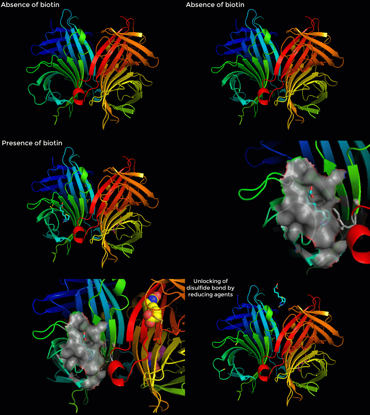
The research group found success recently when they combined molecular design with previous knowledge on the streptavidin-biotin system to discover the effects of newly introduced disulfide bonds, one of which improved the strength of the bond while also creating a reversible switch. Using the CMCF beamline at the Canadian Light Source (CLS) at the University of Saskatchewan, they analyzed the effects of these changes on the structure of the streptavidin-biotin complex and published their findings in Scientific Reports.
“It’s not a perfect system yet, but I think it’s the first time that someone has created a switch like this where you can use something mild to break apart the bond without weakening the tight binding that is so desirable in this system,” Ng said.

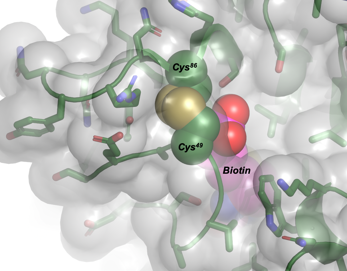
The new discovery involves creating specific bridges that are created by two residues of the amino acid cysteine. The trick was to place the bridges, known as disulfide bonds, at just the right places in the streptavidin structure to alter the binding between streptavidin and biotin.
“By creating a disulfide bond at just the right place in the structure, which is the novel discovery in this work, you can make the binding even tighter.” Ng said, “The cool thing is that when you reduce the disulfide bond the binding of biotin to streptavidin becomes much weaker. Like a switch, the looser binding allows for streptavidin-biotin to come apart, which makes it reversible and solves the biggest problem for the system.”
Many diagnostic tests and biotechnological applications, like ligand capture, protein immobilization and western blotting, prefer a tight bond that leaves behind something permanent. If you are detecting bacteria or viruses using a biosensor you want that bond to be tight so the probe doesn’t slip off.
Medical applications, such as stem-cell therapy used as an experimental treatment for a growing list of ailments, often need something that is reversible. A tight bond is needed to grab and hold on to another protein or cell, but having a way to release it is just as important, especially if you are working with delicate, living cells that are being used in medical treatments.
Using the CLS, the team compared the effects of two different disulfide bonds. The structures were crucial for understanding how the introduction of different mutations affected the strength and stability of the modified forms of streptavidin with biotin.
The team was surprised to discover that one alteration led to a much weaker interaction — the opposite of what was intended. With the help of the CLS, the group was able to visualize the results, which helped to inform their design and push them closer to a perfect system.
Marangoni, Jesse M., Sau-Ching Wu, Dawson Fogen, Sui-Lam Wong, and Kenneth KS Ng. "Engineering a disulfide-gated switch in streptavidin enables reversible binding without sacrificing binding affinity." Scientific reports 10, no. 1 (2020): 1-15. DOI: 10.1038/s41598-020-69357-5.
For more information, contact:
Victoria Schramm
Communications Coordinator
Canadian Light Source
306-657-3516
victoria.schramm@lightsource.ca
