Dairy discovery could improve dialysis design for kidney failure patients
Researchers with USask Engineering were able to view an industrial milk-filtering #membrane in a way not seen before using our BMIT beamline
By Colleen MacPhersonPouring milk on black background

Researchers were able to view an industrial milk-filtering membrane in a way not seen before – layer by layer at a microscopic scale, as skim milk flowed through. Their observations hold great promise for increased yields and creating new dairy products, and could even help in redesigning hemodialysis membranes, and thus patients requiring kidney dialysis treatment.
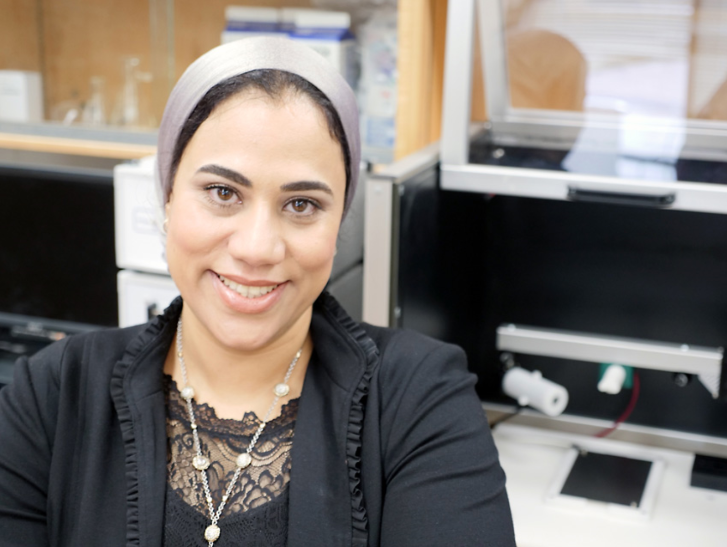
“When you realize your key findings will help patients, you get a different feeling than when you’re working with a single industry,” said Dr. Amira Abdelrasoul, assistant professor with the College of Engineering at the University of Saskatchewan and lead of the university’s membrane science and nanotechnology team.
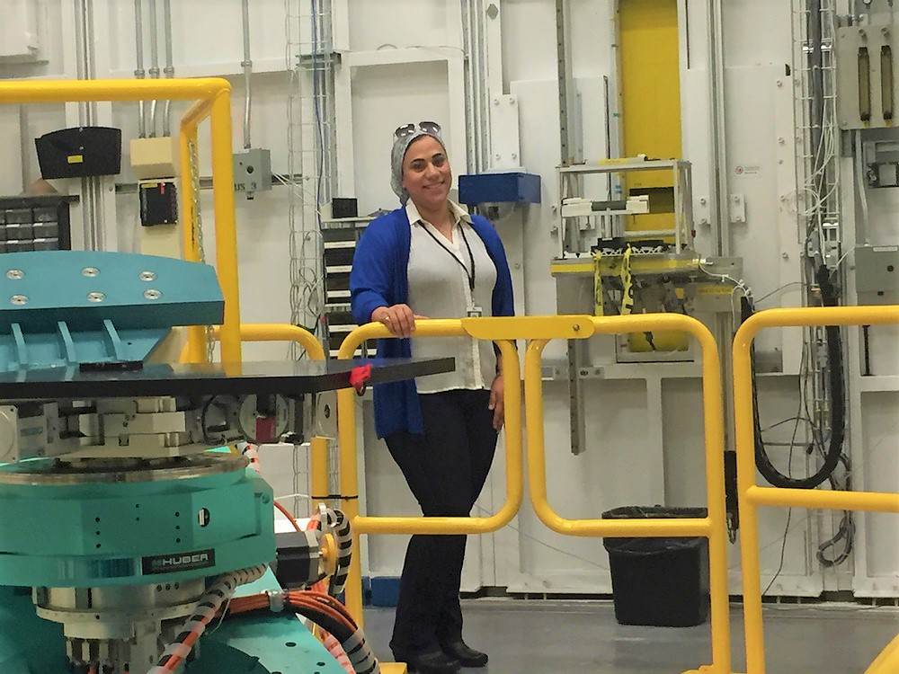
Her group used synchrotron light millions of times brighter than the sun at the Canadian Light Source (CLS) at the University of Saskatchewan to get this unprecedented view at the membrane, the results of which were published online. They used the facility’s BMIT beamline to create images of ultra-thin, multi-layered ceramic membranes used to filter milk. The goal was to determine why and where they clog up, or foul, in the process.
“What we saw was milk protein components deposited in the membrane pores,” said Abdelrasoul. “Other imaging techniques have only allowed us to see the top layer of the membrane but synchrotron techniques allow us to see each layer of these very thin membranes, which means we can assess the protein deposition and fouling at all points in the process.”

In the dairy industry, fouling is a major concern, she said. “In the filtration process, separating out proteins to create other products can clog membranes, which requires a higher energy demand, reduces the membrane lifespan and increases cleaning frequency, and these are all costs to the industry. The same thing happens when dairy proteins are filtered out of waste water for other applications.”
In addition to being able to see where membrane clogging takes place, the images produced on the BMIT beamlines will allow Abdelrasoul to model fouling under different operating conditions. This opens the door to predicting optimal design and operating conditions for various membranes structures.
After this discovery, Abdelrasoul quickly returned to the focus of her research program– hemodialysis, a blood filtration process used to treat kidney disease.
“The imaging we were able to do at the CLS was the tool I was looking for, not just for ceramic membranes used in the dairy industry but also for the polymer membranes used in hemodialysis, where fouling can trigger inflammation in dialysis patients, due to protein deposits in the filtration membrane.”
Being able to see where, when, and how fouling occurs offers the opportunity to use CLS technology to design and assess membranes that will operate efficiently and effectively for both the dairy industry and human health-care.
“I’ve never seen inside a membrane before,” said Abdelrasoul, “so the synchrotron is my magic tool.”
Written by Colleen MacPherson. Photos: Synchrotron | BMIT
To arrange an interview, contact:
Victoria Schramm
Communications Coordinator
Canadian Light Source
306-657-3516
victoria.schramm@lightsource.ca
